|
Estimated results
The research results will be published in high impact impact journals or will be presented in national and international scientific events. Annual reports as well as the final report will be presented.
The research results: PDF version 
Results on October1st, 2019
I. Summary
All the activities proposed to be carried out during the period from June 2017 to October 2019 of the project "Conformational modifications of peptides in the presence of metal ions and anti-amyloid compounds, dependent on time and pH, involved in neurodegenerative diseases" were fully accomplished. In phase I, in 2017, an HPLC instrument was purchased. Training of the personnel involved in the project, including the PhD students, was carried out. The documentation and training of the project members with the related activities were carried out, the specific methods of working in the laboratory were established, using the appropriate tools available and the data obtained were processed. In this first step, some amyloid peptides such as A(4-10), A(1-10), A(1-28) were synthesized, which were characterized by mass spectrometry, infrared spectroscopy, circular dichroism, atomic force microscopy, and spectrophotometry. Among the new neuroprotective peptides (called anti-amyloid), analogues of cysteine and alanine of the NAP peptide were synthesized. In the second year 2018, new amyloid peptide fragments derived from Aβ(9-16) were synthesized, such as peptides in which the tyrosine residue at position 10 was replaced with glycine or phenylalanine, as well as those in which the residues or one of the histidine residues were replaced with other amino acid residues. These were characterized by mass spectrometry, infrared and circular dichroism spectroscopy, atomic force microscopy, spectrophotometry and other methods. Among the anti-amyloid peptides, in addition to the cysteine and serine analogues of the NAP peptide (H2N -NAPVSIPQ-COOH), other peptides, for example, have been synthesized, for example: NAPG (H2N-NAPVGIPQ-COOH), glycine analog, NAPA (H2N - NAPVAIPQ-COOH), analogous to alanine, NAPH (H2N -NAPVHIPQ-COOH), analogous to histidine NAPY (H2N -NAPVYIPQ-COOH), tyrosine analog, and Asp-NAP (Asp-HN-NAPVSIPQ-COOH), a conjugate of NAP peptide with acetylsalicylic acid.
In 2019, the third stage, the interactions of the amyloid peptides with the neuroprotective ones, as well as with other antiaggregant compounds, such as resveratrol, curcumin, etc. were studied. Some results have already been presented at the international conference SGEM2019, Albena, Bulgaria.
Two mobilities were made at the University of Chemical Technology and Metallurgy in Sofia, Bulgaria, and some of the project members participated in two editions of the Summer School in the field of biochemistry and mass spectrometry, IRO Iasi.
The dissemination of the results was also made by presenting a number of 11 (eleven) scientific papers and contributions, orally or as posters at international and national conferences. A number of 15 (fifteen) scientific papers were drafted mentioning the financing of the project. Then, 8 (eight) papers have already been published in impact factor journals. Other two papers have been accepted for publication, and one has been published first on line. Other 4 (four) papers were published in the volumes of the international conferences ISI quoted. Thus, the results of the project exceeded the expectations of the proposal made in 2017, this being mainly due to the doctoral students who had the thesis topic in the field of this project.
II. Scientific and technical description
Introduction
We started from the idea that Alzheimer's disease (AD) is a major cause of disability and mortality, being characterized by an insidious decline of memory, which affects language, video space perception, the ability to perform calculations and executive functions. Behavioral changes are common in AD and include psychosis, agitation, depression, anxiety, personality changes and neurovegetative changes.1-3 Diffuse plaques, representing the earliest stage of amyloid peptide deposition, were exclusively positive for peptide A42, but completely negative for peptide A40.4 In AD, amyloid Aβ oligomers negatively affect synaptic structure and plasticity. Findings from other neurodegenerative diseases indicate that a process similar to neuronal dysfunction is induced by oligomers of modified conformational proteins diffusing into the brain.5 It has also been suggested that Aβ accumulation also affects memory independent of plaque or neuronal loss and this peptide may contribute to the cognitive impairments associated with Alzheimer's disease.6
Our research in this project concerns two distinct lines of investigation: amyloid peptides, derived from amyloid precursor protein (APP) 7, and anti-amyloid (neuroprotective) peptides derived from the NAP peptide, which prevent the formation of toxic oligomers. NAP is an eight-amino acid neuroprotective peptide (NAPVSIPQ), derived from activity-dependent neuroprotective protein (ADNP) responsible for gene regulation and brain development.8 Octapeptide discovery is linked to known vasoactive intestinal peptide (VIP) studies for their neuroprotective activities.9 NAPVSIPQ interacts with microtubules to ensure strong neuroprotection, reduce your hyperphosphorylation, and reduce the decrease of amyloid peptide aggregation. AD disease is correlated with the two prolines found in its primary structure (Asn-Ala-Pro-Val-Ser-Gln).10
From the amyloid peptides we selected to start this project the fragments A(1-10) and A(4-10) that we used in the study of the binding of copper and zinc ions. Next, we studied A(1-16) peptide and its analogs, which have the ability to bind metal ions, such as iron, copper, zinc and even aluminum, as well as A(9-16) fragment peptide and its analogues, since we suspect that this sequence would be heavily involved in metal binding.
Two PhD students have been directly involved in this project, who have already finished their theses: "Anti-amyloid peptides: synthesis, characterization, interaction with other biologically active peptides" (Chemist Lupăescu Ancuţa-Veronica) and, respectively, "Amyloid peptides: synthesis, characterization and biomedical applications” (Chemist Monica Jureschi-Iavorschi). These works will be defended in November 2019 and February 2020, respectively.
Degree of achievement of the objectives
II.2.1. Activity 1.1. Updating information on mass spectrometry and molecular systems involved in the degenerative diseases neurochemistry-permanent
Degree of achievement of the objectives
All scientific information was updated and the project team members were further trained in mass spectrometry. Practically, new various substances were tested in order to establish the suitable matrix for our investigated peptides. Moreover, some screening assays using some amyloid and anti-amyloid (NAP type) peptides have been made, establishing the influence various heavy metals on peptide folding and aggregation. A set of in vitro experiments with our newly synthesized peptides was done. Manual peptide synthesis of peptides such as NAP-OH, NAP(S→C), A(4-10) and A(1-10) was preferred instead of automatic one. One of the main reasons was to give PhD students the opportunity to understand in detail the entire strategy of solid phase synthesis.
In the present project, we performed the synthesis and characterization of two NAP-like peptides (H2N-NAPVSIPQ-OH and its Cys5 mutant). Supplementary, their copper (I) and silver complexes were investigated in detail. The binding of heavy metal ions to NAP peptides was investigated after long-term exposure of the peptide to relatively high concentrations of Cu and Ag ions. Afterwards, all investigated peptide and their peptide-metal ion mixtures were analyzed by mass spectrometry using a MALDI-TOF instrument. The binding of the metal to the peptides was also confirmed by FTIR spectroscopy, which revealed significant changes in the peptide secondary structure.
MALDI-ToF mass spectra of (a) NAP peptides (NAPVSIPQ) and (b) its mutant (NAPVCIPQ).
The mass of the peptides [M], but also of their fragments at M-17 (loss of NH3 in the spectrometer under the action of the laser at 337 nm), as well as some fragments left attached to the peptides are observed. In addition, in the following mass spectra the complexes of these peptides can be detected with silver ions, which appear differently depending on the peptide investigated.
MALDI-ToF mass spectra of native NAP peptide (NAPVSIPQ) and its mutant (NAPVCIPQ) in the presence of silver ions (1: 1 molar ratio). a) NAP plus Ag+ b) NAPCOH plus Ag+.
II.2.2. Activity 1.2. Establishing working methodology and the data processing methods
The methodology was established and the project members were trained on the project activities. Then, the specific methods of working in the laboratory were established, using the appropriate instruments available and the data obtained were processed.
In addition to these activities, the binding of copper ions to some newly synthesized peptides has been studied. These investigations are in progress and a work on the copper binding to fragment A(4-10) is in progress. Thus, the copper ions bind to the amyloid peptides, A(1-40) and A(1-42), causing the modification of their conformation from random coil to -sheet, with oligomerization and fibrillation. However, binding is made in the N-terminal region of these peptides, namely to the fragment A(1-16) which is also the water soluble part of these molecules. But, it is not known exactly which amino acid residues the metal ions bind to, although many researches carried out with various techniques such as electrochemical, spectrophotometric, FT-IR, etc. In this project, we synthesized the peptides A(4-10) and A(1-10), respectively, we purified and characterized them by mass spectrometry, ESI-ion trap-MS.
We proceeded to bind copper and zinc ions, at pH 7.4, and to peptide molar ratios: 1: 1 metal ions; 1: 2 and 1:10, respectively. We performed both MS and MS/MS spectra to study peptide fragmentation and metal complex formation with both peptides and their fragments. In this way, the binding site of the metal ions can be determined precisely and a correct mechanism of binding of these ions can be proposed.
MS/MS spectrum of amyloid peptide fragment A(4-10), which proves its structure.
Mass spectrometric measurement showed the binding of a single copper ion to the peptide fragment A(4-10), while peptide A(1-10) binds less than one copper ion, but its binding is dependent the concentration.
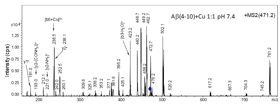
MS/MS spectrum of peptide complex A(4-10) with copper ions.
II.2.3. Activity 1.3. Purchase equipment (HPLC instrument), materials, reagents, consumables, glassware necessary to conduct the proposed activities
A HPLC instrument was purchased, training was carried out for the personnel involved in the project, including doctoral students. The documentation and training of the project members with the related activities were carried out, the specific methods of working in the laboratory were established, using the appropriate tools available and the data obtained were processed.
II.2.4. Activity 1.4. Creation Web site of the project
The web page has been created, but the main results will be published after the completion of this report.
II.2.5. Activity 2.1. Chemical synthesis and characterization of beta-amyloid peptides and their complexes with metal ions at different pH values and concentrations by MS, CD, FT-IR, NMR, AFM and SEM
Stage 2 - 2018 had as a general objective "the synthesis and characterization of mutant peptides and beta-amyloid complexes". In principle, to understand the physiological properties and role of peptides involved in neurodegenerative diseases, similar peptides are being used, but in which some amino acids are replaced by other amino acids, thus altering the characteristics of the original peptides. Mutant peptides are obtained with new properties, which can help to understand the role of peptides in the living cell, in general. On the other hand, normal peptides, such as those involved in Alzheimer's disease, interact with metal ions which are found in brains in physiological concentrations to undergo conformational changes that allow them to aggregate and form fibrils. These fibers form amyloid plaques that include such metals as copper, iron and zinc. The literature offers numerous examples of such structures, in which the metal ions play the role of redox systems that lead to the formation of free radicals (ROS) with potentially destructive neurons.11 Although aging is considered a risk factor for Alzheimer's disease (AD), substantial evidence suggests that an imbalance of essential biometal ions in the body and exposure to certain metal ions in the environment may cause AD disease changes.
The researches continued with the solid phase synthesis of new peptides, mutants of amyloid peptides, as well as of NAP-like, neuroprotective ones, using the methods described in the previous report, of Stage I. In the second stage, new complexes of the newly synthesized peptides were obtained being characterized with modern methods. We have thus benefited from the support of the Iaşi Regional Institute of Oncology, which has a MALDI-TOF spectrometer and colleagues from the University of Chemical Technology and Metallurgy of Sofia, where it is an ICP-OES spectrometer. In this second stage of the project, there were synthesized, besides the native fragment Aβ(9-16) GYEVHHQK, which is necessary for comparison, two other mutant fragments Aβ(9-16)Y10G and Aβ(9-16)Y10F that are not commercially available. In A(9-16)Y10G and Aβ(9-16)Y10F tyrosine (Tyr, Y) from position 10 was replaced with glycine (Gly, G) and phenylalanine (Phe, F), respectively. The synthesis of amyloid peptide fragments was performed by the solid phase manual method using the Fmoc/t-Bu strategy.14,15 Coupling was performed on two types of resin. Aβ(9-16) fragments have been synthesized on a Fmoc-Rink-Amide type resin with a binding capacity of 0.48 mmol amino acid/g resin.
Five amyloid peptides and five anti-amyloid peptides were synthesized in solid phase on the polymeric resin. The synthesis of amyloid and neuroprotective peptide fragments was performed by the solid phase manual method using the Fmoc/t-Bu strategy. For coupling the Aβ(9-16) fragments a Fmoc-Rink-Amide type resin with a binding capacity of 0.48 mmol amino acid/g resin was used. In the case of NAP-type peptides, the synthesis was performed on an Fmoc-Gln (Trt) -Wang Resin resin with a binding capacity of 0.4-0.8 mmol amino acid/g resin.
Manual solid phase synthesis of the peptides was performed in a plastic syringe provided with frit. The synthesis device requires the connection to a vacuum pump in order to remove the washing solutions. Peptide synthesis was performed in DMF (dimethylformamide) medium. Protective groups such as Fmoc or tert-Butyl, used to protect amino acids at the N-terminus and side chain, were removed with 20% piperidine in dimethylformamide.
The precipitation of peptides was carried out cold (-20 ° C) in a solution of ethyl ether. Collecting the fractions required the preparation of a filtration system consisting of a conical filtration glass, a funnel with a fry and a rubber stopper. Peptide extraction was performed with 5% acetic acid solution. Fractions collected in Eppendorf vials were lyophilized and weighed.16,17 In the case of peptides of the type Aβ (9-16), besides the native fragment Aβ (9-16), necessary for comparison GYEVHHQK, since it is not commercially available, four mutant fragments were obtained. By replacing tyrosine (Tyr, Y) at position 10 with glycine (Gly, G) and phenylalanine (Phe, F), respectively, the following peptides were obtained: Aβ(9-16)Y10G: GGEVHHQK and Aβ(9-16)Y10F: GFEVHHQK. Also, two fragments were synthesized in which the two histidines (His, H) from positions 13 and 14 were replaced with glycine (Gly, G) and a fragment in which the tyrosine (Tyr, Y) was replaced from the position 10 with glycine (Gly, G) respectively, the two histidines (His, H) at positions 13 and 14 with glycine (Gly, G), the following peptides being obtained: Aβ(9-16)H13GH14G: GYEVGGQK and Aβ(9-16)Y10GH13GH14G: GGEVGGQK.
In the case of NAP-type anti-amyloid (neuroprotective) peptides (NAP = NAPVSIPQ), these syntheses targeted the SIP (Ser-Ile-Pro) sequence of the native peptide considered by some researchers to be its active center (Oz et al. 2014). By replacing the serine with alanine, glycine, histidine or tyrosine, mutant peptides were obtained that could have therapeutic properties in neurodegenerative diseases. Mass spectrometry allows the determination of molecular mass, purity and structural elements. Comparing the theoretical m/z values with the signals obtained practically in the mass spectrum, it was possible to confirm the obtaining of the desired compounds.
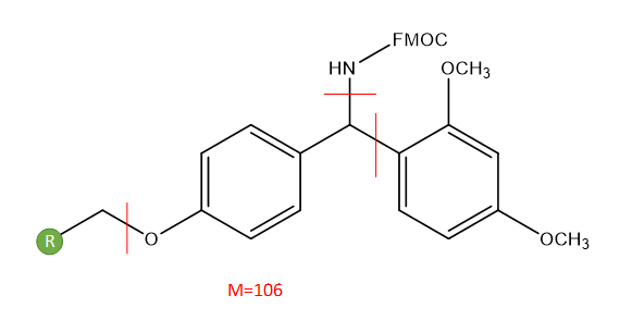
The mass spectrum confirmed the peptide synthesis (Fig. 1). The characteristic signal of the molecular ion [M+H]+ was observed at m/z 996.780, and corresponds to the theoretically calculated value, where [M+H]+ is found at m/z 996.075. In the spectrum are there the signals attributed to the adducts of the peptide with Na+ and K+: [M+Na]+ at m / z 1018.781, and [M+K]+ at m/z 1034.760. The characteristic signals of the unprotected peptide [M+H+106]+ and of its adducts with [M+Na+106]+ and [M+K+106]+ were also observed. The protective group with mass 106 Da represents a residue of a real protective group, with the formula below:
As for peptide Aβ(9-16)Y10G, the molecular ion [M+H]+ is at m/z 890.696, the [M+Na]+ ion, at m/z 912.690, and the signal for [M+K]+ at m/z 928.666 in the MS spectrum (Fig. 2). The experimental values are very close to theoretically calculated values, in which the molecular ion [M+H]+ is found at m/z 890.460, [M+Na]+ ion at m/z 912.442 and [M+K]+ at m/z 928.416. Au fost găsite semnale datorate peptidei incomplet deprotejată și aducții şi [M+K+106]+. Signals due to the incompletely deprotected peptide [M+H+106]+ and adducts [M+Na+106]+ and [M+K+106]+ were found as well. At m/z 1104.837 the signal corresponding to the matrix was identified.

Fig. 1. The MALDI-TOF mass spectrum in a positive reflectron of Aβ(9-16) peptide.
In addition, the phenylalanine-modified Aβ(9-16) peptide sequence (Fig. 3) was confirmed by mass spectrometry. Thus, the molecular ion [M+H]+ was found at m/z 981.786, and the [M+Na]+ ion at m/z 1003.772. The values obtained were practically close to the values calculated theoretically, in which the molecular ion [M+H]+ was at m/z 890,411, while the [M+Na]+ ion was found at m/z 1003.065. At the same time, at m/z 1194.95, the signal corresponding to the matrix was observed, with some signals being attributed to the incompletely deprotected peptide (Fig. 3).
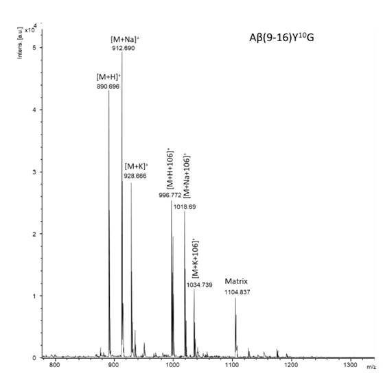
Fig. 2. MALDI-ToF mass spectrum in a positive reflectron of the modified peptide Aβ(9-16)Y10G.
Purification of amyloid peptides was performed using high performance liquid chromatography (HPLC). The mobile phase used consisted of a mixture of: solvent A (0.1% TFA) and solvent B (80% ACN in 0.1% TFA). The separation of hydrophilic peptides of the type Aβ(9-16) was performed on an analytical Vydac C18 column using the gradient presented in Table 1. Monitoring of the peptides was performed at λ 214 nm, characteristic wavelength, at which the peptides absorb. Thus, in the chromatogram of the peptide Aβ (9-16) (Fig. 4.) two well separated signals appear.
After purification, the mass spectrum confirmed the obtaining of the Aβ peptide (9-16) by the characteristic signal of the molecular ion at m/z 996.855 which is in agreement with the theoretically calculated value, where [M+H]+ is found at m/z 996.075 (Fig. 5). In the spectrum, the signals of the adducts with Na+ and K+ are also present. Also present are signals due to the incompletely protected peptide [M+H+106]+ and its adducts [M+Na+106]+, [M+K+106]+.
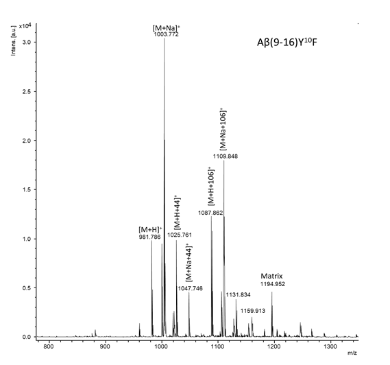
Fig. 3. MALDI-ToF mass spectrum in a positive reflectron of the modified peptide Aβ(9-16)Y10F.
The mass spectrum of glycine-modified Aβ(9-16) peptide instead of the two histidines confirmed that the peptide was successfully obtained (Fig. 4). Basically, the molecular ion [M+H]+ was at m/z 836.753, the [M+Na]+ ion, at m/z 858.744, and the signal for [M+K]+ at m/z 874.716. The experimental values were very close to the values calculated theoretically, in which the molecular ion [M+H]+ is found at m/z 836.408, the ion [M+Na]+ at m/z 858.397, and the ion [M+K]+ at m/z 874.506. Also, at m/z 942.834 and at m/z 1058.909 the signals corresponding to the incompletely deprotected peptide were observed.
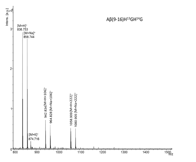
Fig. 4. MALDI-ToF mass spectrum in a positive reflectron of the modified peptide Aβ(9-16)H13GH14G.
The glycine-modified peptide Aβ(9-16) was synthesized at positions 10, 13 and 15, and the molecular mass of this peptide was confirmed by MALDI-ToF mass spectrometry (Fig. 5). The molecular ion [M+H]+ was found at m/z 730.680, the [M+Na]+ ion, at m/z 752.670, and the characteristic signal of the [M+K]+ ion at m/z 768.760. The experimental values are very close to the values calculated theoretically, in which the molecular ion [M+H]+ is at m/z 730.366, while the ion [M+Na]+ at m/z 752.355, and [M+K]+ at m/z 768.464. Signals attributed to the incompletely deprotected peptide [M+H+106]+ and [M+H+222]+, as well as their adducts [M+Na+106]+, [M+Na+222]+, were also identified.
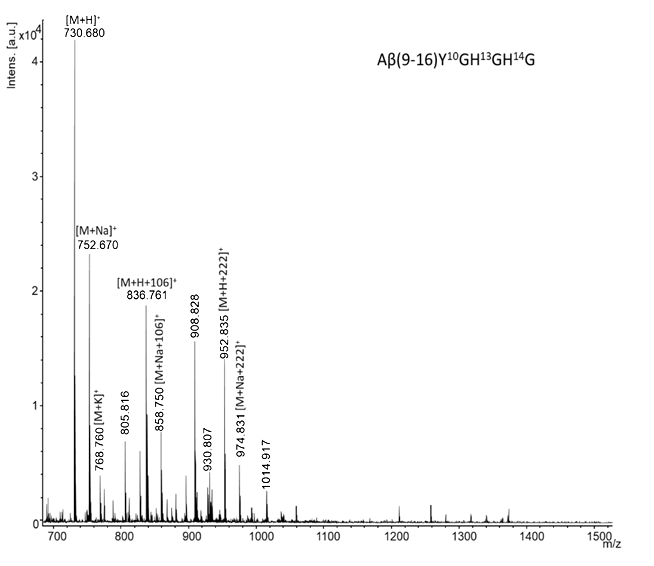
Fig. 5. MALDI-ToF mass spectrum in a positive reflectron of the modified peptide Aβ(9-16)Y10GH13GH14G.
Thus, in the chromatogram of the peptide Aβ(9-16) (Fig. 6.) two well separated signals appear, one at minute 13.70, which is corresponding to the pure peptide and the second at minute 18.83 corresponding to the protected peptide. This is also confirmed by mass spectrometry.
After purification, the mass spectrum confirmed the obtaining of the Aβ(9-16) peptide by the characteristic signal of the molecular ion at m/z 996.855 which is in agreement with the theoretically calculated value, where [M+H]+ is found at m/z 996.075 (Fig. 7). In the spectrum, the signals of the adducts with Na+ and K+ are also present. Signals due to the incompletely protected peptide [M+H+106]+ and its adducts [M+Na+106]+, [M+K+106]+ are present as well.
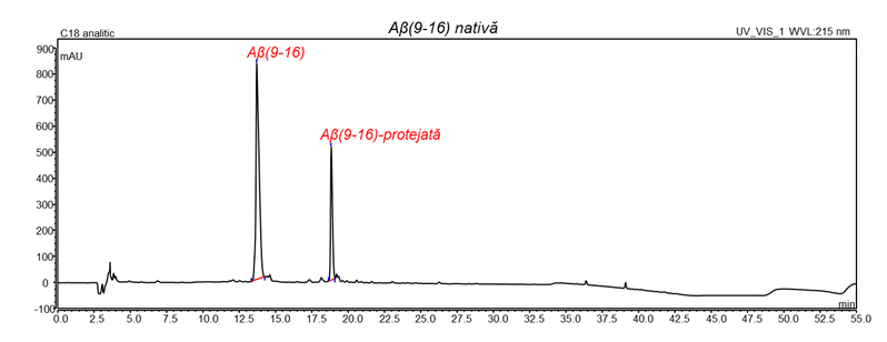
Fig. 6. Chromatographic separation of the peptide Aβ(9-16).
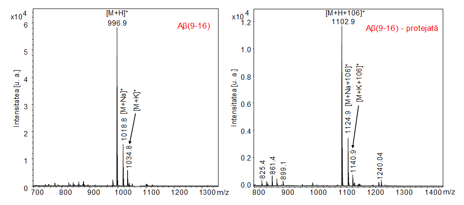
Fig. 7. The MALDI-TOF mass spectrum in positive reflectron mode of the Aβ(9-16) peptide after purification, as well as that of the partially protected peptide.
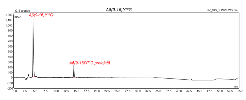
Fig. 8. Chromatographic separation of the peptide Aβ(9-16)Y10G (protected = protejată).
In the chromatogram of peptide Aβ(9-16)Y10G (Fig. 8.) two well separated signals were observed, one at minute 4.663, corresponding to the pure peptide and the second signal at minute 14.643, assigned to the protected peptide.
After separation, the mass spectra confirmed the obtaining of pure peptide, the molecular ion [M+H]+ being at m/z 890,811, which is in agreement with the theoretically calculated value. In the spectrum, the signals of the adducts with Na+ and K+ are also present (Fig. 9.a). In Fig. 9.b the signals of the incompletely deprotected peptide [M + H + 106]+ and its adducts [M + Na + 106]+ and [M + K + 106]+ are present.
Fig. 8. Chromatographic separation of the peptide Aβ(9-16)Y10G (protected = protejată).
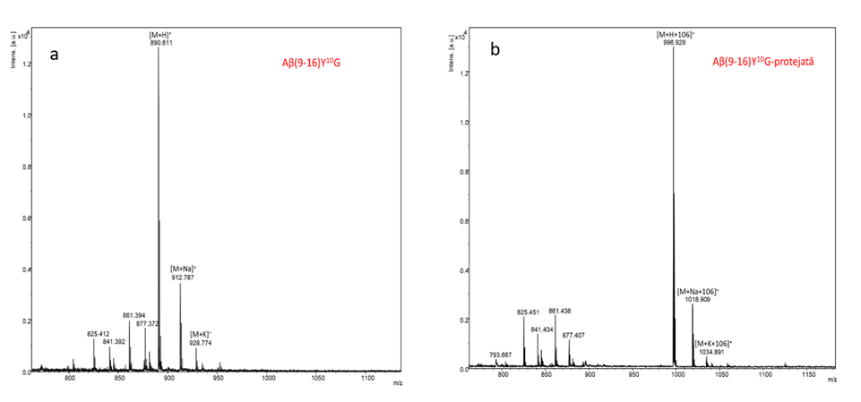
Fig. 9.(a,b) MALDI-ToF mass spectra in positive reflectron mode of Aβ(9-16)Y10G peptide after purification.
In Fig. 10 the chromatogram of the peptide Aβ (9-16) Y10F is shown in which two well-separated peaks are observed, at the retention time of 18,527 (pure peptide) and a second peak at the retention time 22,790 corresponding to the protected peptide.
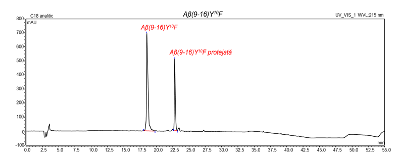
Fig. 10. Chromatographic separation of Aβ(9-16)Y10F peptide (pure peptide and protected one, protejata).
After purification, the synthesis of Aβ(9-16)Y10F peptide was proved using MALDI-Tof mass spectrometry due to the characteristic signal of the molecular ion [M+H]+ from m/z 981,890, which is very close to the theoretically calculated value. In this spectrum, the signals of the adducts with Na+ and K+ were also present (Fig. 11.a). The next figure shows the signals due to the incompletely protected peptide [M+H+106]+ and the respective [M+Na+106]+ and [M+K+106]+ respectively (Fig. 11.b).
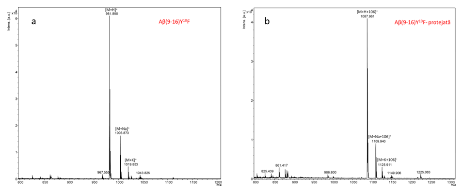
Fig.11.(a,b) MALDI-ToF mass spectra in a positron reflectron mode of Aβ(9-16)Y10F peptide after purification (protected = protejata).
In the chromatogram obtained for the peptide Aβ(9-16)H13GH14G (Fig. 12.) two well-separated peaks were observed, one at the retention time 14.890 min (the pure peptide) and the second at the retention time 20.863 , corresponding to the protected peptide.
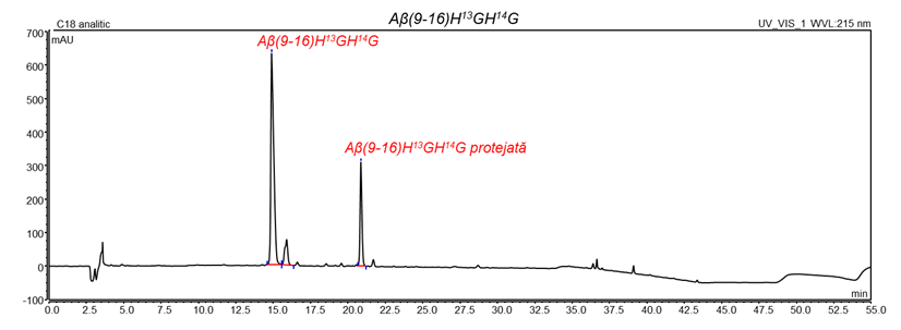
Fig. 12. Chromatographic separation of the peptide Aβ(9-16)H13GH14G.
Following the separation of the pure peptide from the protected one, the mass spectrum of the pure peptide showed the molecular ion [M+H]+ at m/z 836.756, very close to the theoretically calculated value [M+H]+ of m/z 836.408, at a very high resolution of (836,756-836,408) / 836,408 = 0.041%. In the spectrum of pure peptide, the signals of the adducts with Na+ and K+ were also observed (Fig. 13.a). In the adjacent spectrum (Fig. 13.b) the signals of the incompletely deprotected peptide [M+H+106]+, as well as its adducts [M+Na+106]+ and [M+K+106], were present.
The chromatogram of the peptide Aβ(9-16)Y10GH13G H14G is presented in Fig. 14 in which the two well-separated peaks can be observed at the retention time of 4.910 min, corresponding to the pure peptide and a second peak respectively at the retention 15,950 min, corresponding to the protected peptide.
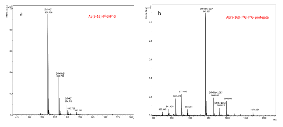
Fig. 13.(a,b) MALDI-ToF mass spectra in positive reflectron mode of the peptide Aβ(9-16)H13GH14G after purification.
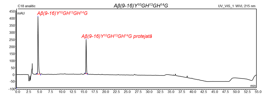
Fig. 14. Chromatographic separation of the peptide Aβ(9-16)Y10GH13G H14G.
After separation, the mass spectra confirmed both the obtaining of the pure peptide and the protected one, the molecular ion [M+H]+ being at m/z 730.601. In the first spectrum, the signals of the adducts with Na+ and K+ are also present (Fig. 15.a). In Fig. 15b the signals of the incompletely protected peptide [M + H + 106]+, as well as adducts [M + Na + 106]+ and respectively [M + K + 106]+ are present.
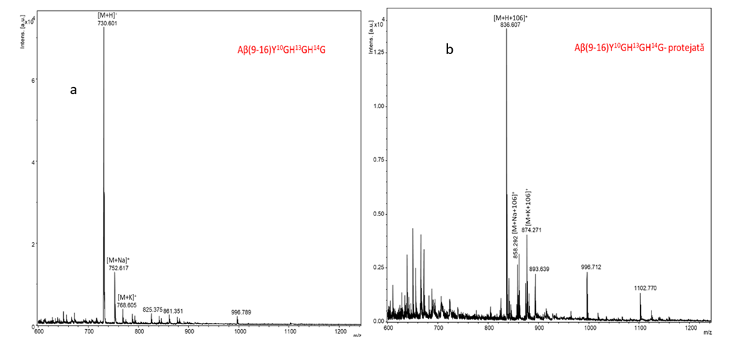
Fig. 15.(a,b) MALDI-ToF mass spectra in positive reflectron mode of the peptide Aβ(9-16) Y10GH13GH14G after purification
The mass spectra of the synthesized peptides can be seen in Figure 16. The NAP peptide showed an intense signal at m/z 847.727 corresponding to the [M+Na]+ ion and a lower signal at m/z 863.702 attributed to the potassium adduct ([M+K]+. The signal at m/z 809.772 corresponding to the [M-16+H]+ ion is formed as a result of the photodecomposition of the peptide under laser power [7]. Other signals corresponding to the partially deprotected peptide were identified at m/z 869.718 ([M+Na]++22) and 1089.922 ([M+Na]++Trt).
The glycyl-mutant peptide showed an intense signal at m/z 779.640 corresponding to the [M-16+H]+ ion. The peptide adducts with sodium and potassium showed signals in the spectrum at m/z 817.614 ([M+Na]+) and 833.741 ([M+K]+), respectively. The peptide showed signals at a difference of 22 units m/z, which cannot be assigned.
The NAPA peptide contains a rather intense signal at m/z 793.831 corresponding to the photodecomposed ion [M-16+H]+. Similar to previous cases, the most intense signals were attributed to sodium (m/z 831.786, [M+Na]+) and potassium (847.766, [M+K]+). The signals at m/z 853.780 and m/z 869.758 contain a difference of m/z 22 relative to the peptide adducts.
The spectrum of the NAPH peptide presents in addition to the ion signal [M-15+H]+ (m/z 859.848) and the molecular ion signal [M+H]+ (m/z 875.851). Other signals were attributed to adducts with sodium (m/z 897.804) and potassium (m/z 913.782). And in this case, signals containing a difference of m/z 242 (Trt) or m/z 22 were found in the spectrum.
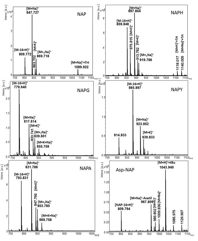
Fig. 16. Mass spectra of the NAP-like peptides: NAP (native), NAPG, NAPA, NAPH, NAPY and Asp-NAP.
In the case of the NAPY peptide, the most intense signal in the spectrum belongs to the ion [M-16+H]+ (m/z 885.897). The signals from m/z 923,852 and 939,833 can be assigned to the [M+Na]+ and [M+K]+ ions. The Asp-NAP conjugate peptide, which represents a conjugate of NAP with acetylsalicylic acid, afforded the most complex spectrum. It contains signals corresponding to the peptide unreacted with aspirin at m/z 809.794, corresponding to the ion [M-16+H]+. A signal of medium intensity was found at m/z 967.809 and was assigned to the [M+Acetyl+Na]+ ion. The molecular ion of the Asp-NAP peptide, [M+H]+, showed a signal at m/z 989.802 while its adduct with sodium ([M+Na]+ was found at m/z 1009.836. The most intense signal in the spectrum (m/z 1043.940) corresponds to the peptide in which the tert-butyl protecting group of the serine remained uncleaved.
The newly synthesized peptides were purified using a Dionex UltiMate 3000 UHPLC chromatographic instrument from Thermo Scientific, Breman, Germany. The separation was performed in an aqueous-organic environment. An aqueous solution of 0.1% TFA (in bidistilled water) (v/v) was used as solvent A, and as solvent B was used 80% ACN, 0.1% TFA in bidistilled water (v/v). All solvents used were of chromatographic purity.
The separation was carried out using a Vydac analytical C18 column of 4.6 mm and 250 mm length. A volume of 250 µl of a testing sample, 0.1 mg/ml concentration dissolved in solvent A, was automatically injected into the chromatographic system. Peptide separation was performed at a flow rate of 1 mL per minute throughout the separation. The UV detector was set at 214 nm, characteristic for peptide groups, throughout the analysis. The working temperature was set to 25˚C.
Table 3. The retention time of the peptides analyzed by RP-HPLC
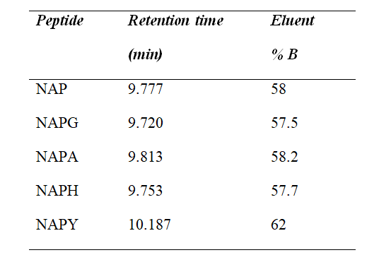
Figures 17–21 show the profile of the chromatograms obtained using the 30 minute gradient. As observed in the resulted spectra, the newly synthesized peptides had a high degree of purity.
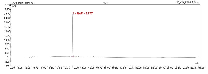
Figura 17 Chromatogram of NAP peptide: NH2-NAPVSIPQ-COOH
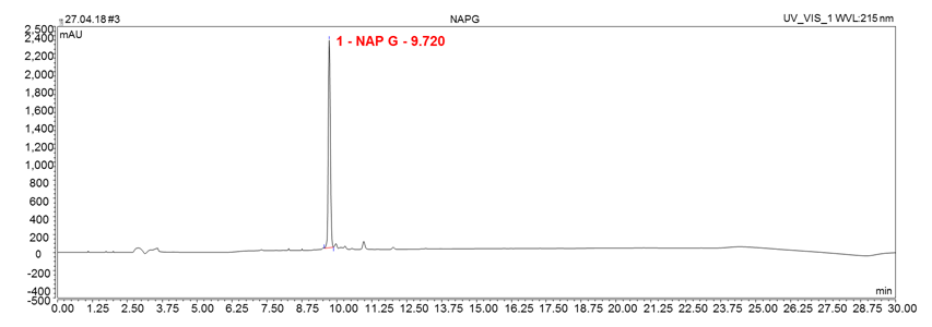
Figura 18 Chromatogram of NAPG peptide: NH2-NAPVGIPQ-COOH.
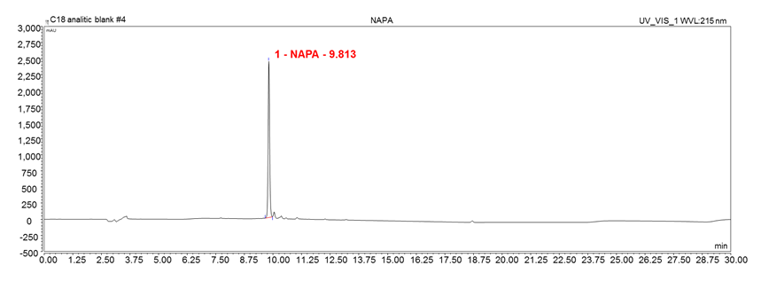
Fig. 19 Chromatogram of NAPA peptide: NH2-NAPVAIPQ-COOH.
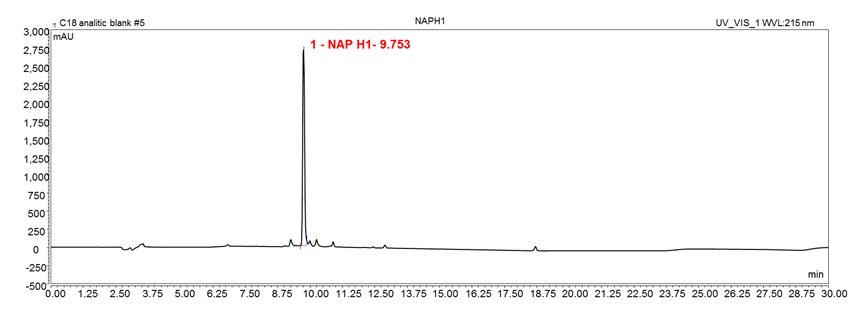
Fig. 20. Chromatogram of NAPH peptide: NH2-NAPVHIPQ-COOH.
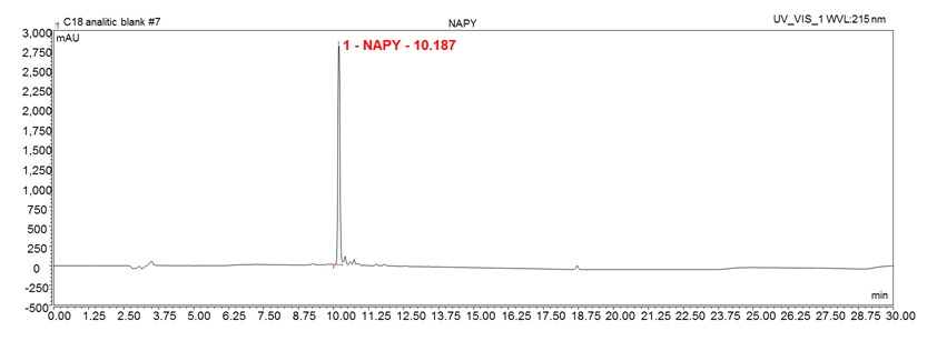
Fig. 21. Chromatogram of the peptide NAPY: NH2-NAPVYIPQ-COOH.
It is observed from this report how the researches were carried out and the results of great significance obtained
II.2.6. Activity 2.2.
Documentation stages abroad on the new techniques for characterization of the studied compounds.
Mobilities were made in November 2018 at the University of Chemical Technology and Metallurgy in Sofia, Bulgaria for the characterization of some metal complexes together with Assoc Prof. Dr. Andriana Surleva and Dr. Darja Ilieva, at the invitation of Bulgarian partners. The field of work has been the measurement of the retention of heavy metal ions in amyloid and neuroprotective peptides. The results obtained show that both types of peptides bind these metals strongly (Fe (II), Ni (II), Zn (II), Cu (II), etc.). Also, some of the members of the project participated in a Summer School in the field of biochemistry and mass spectrometry.
II.2.7. Activity 2.3.
Purchase of equipment, materials, reagents, consumables, glassware.
All the reagents and glassware purchases provided in the project for the year 2018 were made, according to the accounting reports included in the report on this project. These acquisitions were included in the economic report
II.2.8. Activity 2.4.
Peptide synthesis, including beta-amyloid peptides and other necessary compounds
This activity was presented extensively in Activity 2.1, to be short. Although there are some aspects that can be separated between these activities, in principle we introduced them to the same activity. It has already been shown that 5 amyloid peptides and 5 neuroprotective peptides were synthesized during this period, plus their complexes with the transition metals.
II.2.9. Activity 2.5.
The study of chelating agents or anti-aggregating agents
This activity refers to compounds that can chelate heavy metals, such as clioquinol, but we have noticed that NAP-type peptides strongly chelate heavy metal ions and, therefore, we focused on their study. In addition, these peptides have also been shown to be effective antiaggregants. The results can be seen from this report, which presents different complexes between the NAP-type peptides and the multiple metals studied.
II.2.10. Activity 2.6.
Characterization of beta-amyloid peptides and their complexes by HPLC, MS, UV-Viz, FT-IR, AFM, DLS
The newly synthesized peptides were characterized by analytical HPLC, and then separated on a semi-preparative C18 column (Fig. 4). In addition, we noticed a very interesting thing that some peptides cannot be completely deprotected under the working conditions recommended by the literature, but the separation of the pure peptide from the protected peptide can be easily achieved by RP-HPLC, because the two signals in the chromatogram are largely distanced from each other.
All peptides and some complexes were analyzed by UV-vis, and these spectra showed differences between the spectrum sum of the peptide and metal and the spectra of the appropriate mixtures, which denotes a chemical (complex) reaction between species. The same result was obtained by the MALDI-TOF measurements. AFM microphotographs were performed on some peptides and metal complexes. FT-IR studies have shown significant changes in peptide conformation in the presence of zinc, nickel and copper ions. Most of our researches were performed in buffer solutions, and these interfered with the spectral measurements and have to be resumed to highlight only the contribution of the metal ion. DLS studies have started, but only with higher amounts of proteins and peptides in the laboratory. As the dexterity of the operators increases, we will move on to studies on Aβ(1-42) and Aβ(1-40) available in quantities of several milligrams.
Another example of HPLC characterization, separation and then mass spectrometry characterization is shown in Fig. 6. Thus, in the chromatogram of peptide Aβ(9-16)Y10G, two well separated signals were observed, one at minute 4.663, corresponding to the pure peptide and the second signal at minute 14.643, assigned to the protected peptide.
After separation, the mass spectra confirmed the obtaining of the pure peptide, the molecular ion [M+H]+ being at m/z 890.811, which is in agreement with the theoretically calculated value. In the spectrum, the signals of the Na+ and K+ adducts are also present. The signals of the incompletely deprotected peptide [M+H+106]+ and its adducts [M+Na+106]+ and [M+K+106]+ are also present.
II.2.11. Activity 2.7.
Study of the interaction between beta-amyloid peptides, metal ions and chelating agents / anti-aggregates
Part of the synthesized peptides Aβ(9-16), Aβ(9-16)Y10G and Aβ(9-16)Y10F were solubilized in aqueous (degassed) solution at pH 7, close to the physiological (concentration 2 mM buffer) and incubated with Ag+ (Ag2SO4), Cu+ (Cu2O), Cu2+ (CuCl2) and Zn2+ (ZnCl2) ions. The molar ratio peptide: metal ion was 1:10. Only in the case of complexes with silver ions was used a molar ratio peptide: Ag+ of 1: 1, being known in the literature that silver ions have a much higher affinity for peptides than the ions of other heavy metals. The obtained solutions were left overnight stirring in the thermomixer (350 rpm, room temperature). After 24 hours of each sample, 30 µl were collected and mass spectra (MALDI-ToF) were performed.
Thus, in the mass spectrum of the Aβ(9-16):Ag+ complex (Fig. 22), the strong binding of silver ions to the amyloid peptide A(9-16) is observed. The signal of the molecular ion [M+H]+ at m/z 996.822, its adduct with potassium [M+K]+ at m/z 1034.780 and the signal for the silver complex [M+Ag]+ at m/z 1102.742 were identified in the MS spectrum.
Other signals that appeared in the spectrum were noticed at m/z 1159.942, being attributed to the incompletely deprotected peptide [M+H+163]+; at m/z 1210.691 the signal was attributed to the complex with two silver ions [M-H+2Ag]+, at m/z 1265.868 is a signal attributed to the complex between silver and the incompletely deprotected peptide [M+Ag+163]+ and another signal at m/z 1373.815, corresponding to the binding of two silver atoms to the protected peptide [M-H+2Ag+163]+. According to the theoretically calculated values , the signal for the molecular ion [M+H]+ is at m/z 996.075, whereas the complex [M+Ag]+ at m/z 1102.943. The experimental values were very close to the theoretically calculated values .
In the mass spectrum of the Aβ(9-16):Cu+ complex the binding of Cu+ ions to the amyloid peptide can be observed. In this spectrum, the signal of the molecular ion [M+H]+ at m/z 996.852 and the adduct with Na+ [M+Na]+, at m/z 1018.890 were identified. The signal for the Cu+ complex, [M+Cu+]+, was found at m/z 1058.825. The experimental values were very close to the values calculated theoretically, in which the molecular ion [M+H]+ should have been at m/z 996.075, and the [M+Na]+ ion at m/z 1018.065, while the complex ion [M+Cu(I)]+ at m/z 1058.62. In addition, the signals from m/z 1159.953 may be assigned to the incomplete unprotected peptide [M+H+163]+, and the signal from m/z 1221.904 has been attributed to the copper complex of the incomplete deprotected peptide [M+Cu+163]+.
Fig. 22. MALDI-ToF mass spectra of complexes a) Aβ(9-16):Ag+(1:1) și b) Aβ(9-16):Cu+(1:10).
In the mass spectrum obtained for the complex Aβ(9-16):Cu2+(Fig. 23), the reduction of the Cu2+ ion to Cu+ was observed and its binding to the amyloid peptide. Basically, from the MS spectrum we could identify the signals for the molecular ion [M+H]+ which is at m/z 996.81 and the [M+Na]+ ion that is at m/z 1018.83. The signal for the Cu+ [M+Cu(I)]+ ion complex was identified at m/z 1058.75. However, within these measurements, no signal corresponding to the peptide complex: Cu2+ was identified in the mass spectrum. It results in a low affinity for this ion and a higher one for copper would result only after a corresponding reduction. According to the values calculated theoretically, the signal for the molecular ion [M+H]+ was found at m/z 996.075, and the complex [M+Cu(I)]+ at m/z 1058.62. The experimental values were very close to the values theoretically calculated. Signals were also observed at m/z 1159.92, m/z 1181.92 and, m / z 1221.85 respectively, which were attributed to the incompletely deprotected peptide.
In the mass spectrum of the Aβ(9-16):Zn2+ the Zn2+ ion binding to the amyloid peptide can be observed. It was possible to identify the molecular ion [M+H]+ in the spectrum, which is at m/z 996.81 and the adduct with sodium [M+Na]+, at m/z 1018.80. The signal for the complex with Zn2+, [M+Zn2+]+, was at m/z 1059.74. The experimental values were very close to the values calculated theoretically, in which the molecular ion [M+H]+ was at m/z 996.075, and the ion [M+Na]+ at m/z 1018.064; The [M+Zn-H]+ ion was found at m/z 1059.484. Besides, signals from m/z 1124.90 and m/z 1159.92 were assigned to the unprotected peptide.
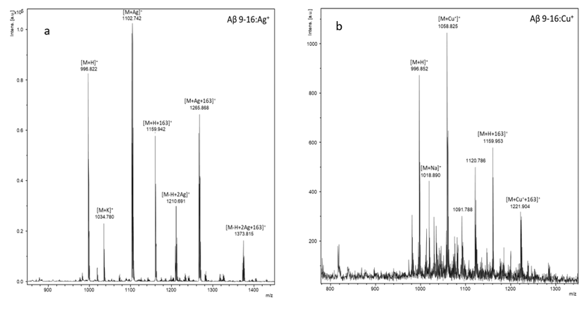
Fig. 23. MALDI-ToF mass spectra în positive reflectron mode of complexes a) Aβ(9-16):Cu2+(1:10) and b) Aβ(9-16):Zn2+(1:10).
These investigations continued with the formation of the Aβ(9-16)Y10G:Ag+ complex. Also in the case of the complex Aβ(9-16)Y10G:Cu+, the binding of Cu+ to the peptide occurred. In the mass spectrum of the Aβ(9-16)Y10G:Zn2+ only the signals of molecular ion [M+H]+ at m/z 890.79 and that of incomplete deprotected peptide at m/z 996.87 were identified. In the case of the phenylalanine-modified Aβ(9-16) peptide complex with Cu2+ ions, it was found that the reduction of Cu2+ to Cu+ occurred, followed by the latter binding to the peptide. Further, multiple interactions are made between amyloid-type peptides and numerous heavy metals with which the organism comes into contact.
Therefore, during this stage, the formation and characterization of complexes of the amyloid peptides of type Aβ(9-16) with different metal ions took place. The newly synthesized peptides Aβ(9-16), Aβ(9-16)Y10G, Aβ(9-16)Y10F, Aβ(9-16)H13GH14G and Aβ(9-16) Y10GH13GH14G were solubilized in aqueous solution (degassed) at a pH 7, close to the physiological one (concentration 4 mM buffer solution) and incubated with Al3+ (AlCl3), Fe3+ (FeCl3), Ni2+ (NiCl2) and Zn2+ ions (ZnCl2). The molar ratio peptide: metal ion was 1:10. The obtained solutions were left overnight stirring in the thermomixer (350 rpm, room temperature). After 24 hours of each sample, 30 µl were collected and mass spectra (MALDI-ToF) were performed. In the mass spectrum of the peptide Aβ(9-16) in the presence of metal ions (Fig. 24), the binding of the Al3+ ion to the amyloid peptide and the formation of the complex was observed; the signal identified at m/z 1020.59. In the case of Fe3+ ions, the reduction of the Fe3+ ion to Fe2+ was observed and its binding to the amyloid peptide; identified signal at m/z 1050.54.
The nickel ion complex can be identified at m/z 1053.41 and the signal for the complex with Zn2+ was at m/z 1059.47. In the case of the peptide Aβ(9-16)Y10G (Fig. 23) the formation of complexes with aluminum (m / z 914.43), iron (m / z 994.46), nickel (m / z 947.44) and zinc (m / z 954.43) was observed from the mass spectra. The mass spectrum obtained for the peptide Aβ(9-16)Y10F (Fig. 24) confirmed the formation of the complexes with aluminum (m/z 1005.49), iron (m/z 1035.47), nickel (m/z 1038,47) and zinc (m/z 1044.50). In the mass spectrum of peptide Aβ(9-16)H13G H14G (Fig. 25) the signals corresponding to the formation of complexes with aluminum (m/z 860.42) iron (m/z 890.52), nickel (m/z 893.50) were identified, but the formation of the complex could not be observed and in the case of zinc ions. Besides, the mass spectrum of the peptide Aβ(9-16)Y10GH13GH14G confirmed the formation of the complexes with aluminum ions (m/z 574.35), iron (m/z 784.41), nickel (m/z 787.53 ) and zinc (m/z 793.32).
The values obtained experimentally were very close to the values calculated theoretically
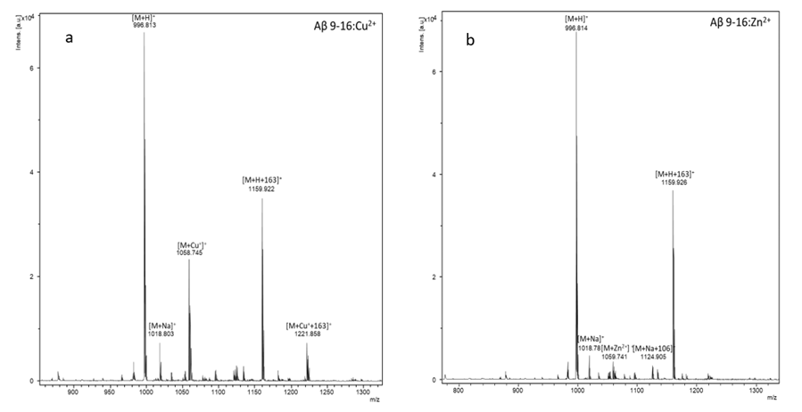
Fig. 24. MALDI-ToF MS spectra of Aβ(9-16) peptide in the absence and presence of Al3+, Fe3+, Ni2+ and Zn2+ ions at a molar ratio of 1:10 peptide: metal ion.
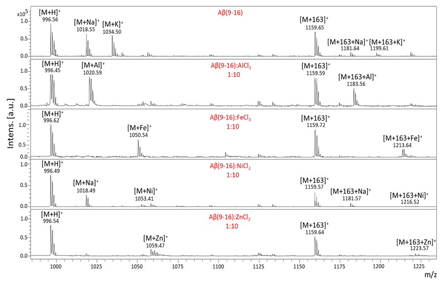
Fig. 25. MALDI-ToF MS spectra of Aβ(9-16)Y10G peptide in the absence and presence of Al3+, Fe3+, Ni2+ and Zn2+ ions at a molar ratio of 1:10 peptide: metal ion.
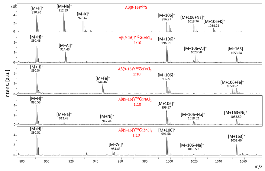
Fig. 26. MALDI-ToF MS spectra of Aβ(9-16)Y10F peptide in the absence and presence of Al3+, Fe3+, Ni2+ and Zn2+ ions at a molar ratio of 1:10 peptide: metal ion.
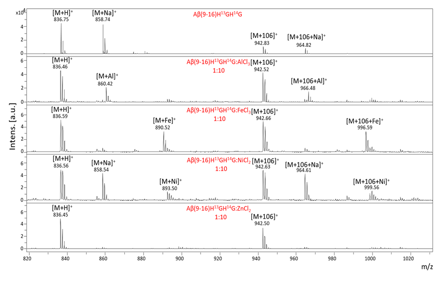
Fig. 27. MALDI-ToF MS spectra of Aβ(9-16) H13GH14G peptide in the absence and presence of Al3+, Fe3+, Ni2+ and Zn2+ ions at a molar ratio of 1:10 peptide: metal ion.
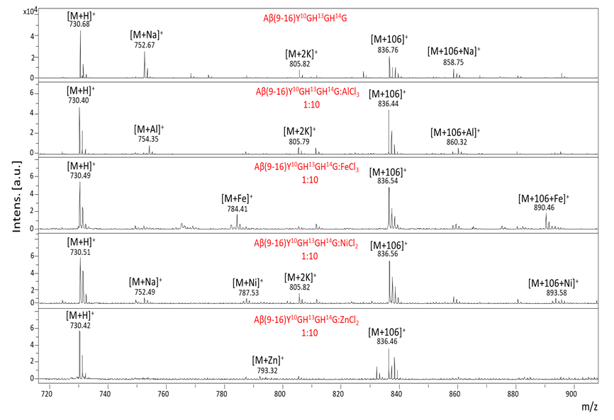
Fig. 28. MS spectra of Aβ(9-16)Y10GH13GH14G peptide in the absence and presence of Al3+, Fe3+, Ni2+ and Zn2+ ions at a molar ratio of 1:10 peptide: metal ion
Binding of heavy metal ions to NAP peptides was investigated following long-term exposure of the peptide to relatively high concentrations of Ni, Zn, and Fe ions. For this purpose, stock solutions of peptides (8 Molar) and metal ion (80 Molar) were prepared in degassed distilled water with pH 7. Following the addition of equal volumes (v/v) of solutions of peptides and metal ion, final solutions were obtained with the molar ratio of 1:10 peptide: metal ion. The samples were stirred (350 rpm) at room temperature for 24 hours. Then, they were co-crystallized in the presence of the HCCA matrix using the dried-drop method.
This technique involves pipetting the sample on the target and then the HCCA supersaturated solution. In order to obtain the crystals, the metallic target containing the sample was left at room temperature. Finally, the sample plate was loaded into the MALDI-TOF MS instrument and analyzed by software. The spectra were recorded with the Bruker Ultraflex MALDI-ToF/ToF mass spectrometer and processed using the flexAnalysis software.
The mass of peptides [M] and their fragments at M-16 (loss of NH3 in the spectrometer under the action of the laser at 337 nm) were identified. In addition, in the following mass spectra the complexes of these peptides can be detected with nickel ([M+Ni]+), zinc ([M+Zn]+) and iron Fe(II) ([M+Fe(II)]+), which appear at different m / z values depending on the peptide analyzed.
In the case of the NAP peptide, following its incubation with different metal ions, the formation of the peptide-metal complexes was observed. As observed in the spectrum below, in the case of Fe ions, the binding of the Fe(II) ion to peptide was observed at m/z 879.59 although the stock solutions were made using FeCl3
Similar results were for the other NAP-type peptides (NAPG, NAPA, NAPH, NAPY). Regarding the stoichiometry of the complexes, it varies depending on the peptide used. If the NAP peptide is capable of complexing two iron molecules with the formation of the [M+2Fe(II)]+ (m/z 933,52) ion, the NAPA peptide has a similar ion in the spectrum corresponding to the nickel sample ([M+2Ni]+ la m/z 921,45). As seen in the mass spectrum of the mutant glycyl peptide, the stoichiometry of the NAPG peptide is similar to the native one ([M+2Fe(II)]+ m/z 903.42) and that of the mutant tyrosine peptide (NAPY) contains as in in the case of the alanine-modified peptide a peak corresponding to the [M+2Ni]+ ion (m/z 1013.46). The mutant histidine peptide (NAPH) is capable of complexing two molecules of nickel, zinc or iron(II), thus demonstrating chelating capacities superior to the other NAP-type peptides.
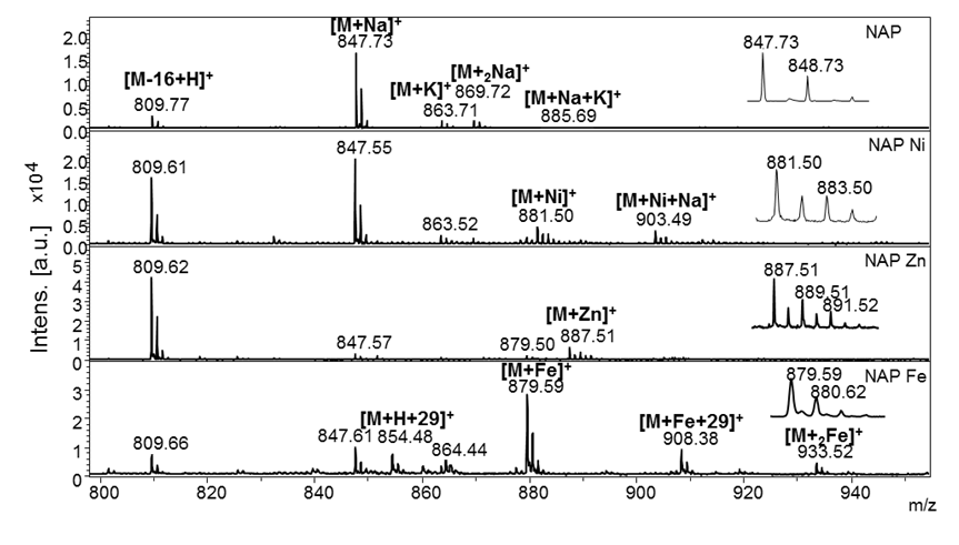
Figura 29. MS spectra of NAP peptide (NH2-NAPVSIPQ-COOH) in the absence and presence of Ni2+, Zn2+ and Fe3+ ions at a molar ratio of 1:10 peptide: metal ion. The insertion in the spectrum corresponds to the isotopic distribution of the peak of interest.
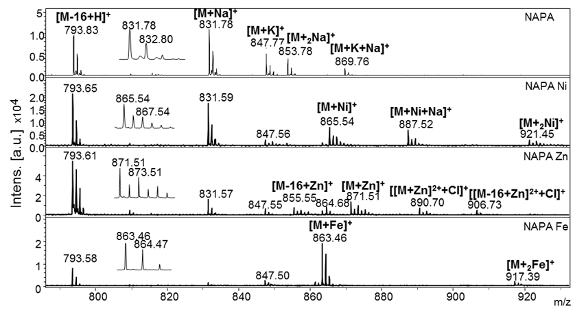
Figura 30. MS spectra of NAPA peptide (NH2-NAPVAIPQ-COOH) in the absence and presence of Ni2+, Zn2+ and Fe3+ ions at a molar ratio of 1:10 peptide: metal ion. The insertion in the spectrum corresponds to the isotopic distribution of the peak of interest.

Figura 31. MS spectra of NAPG peptide (NH2-NAPVGIPQ-COOH) in the absence and presence of Ni2+, Zn2+ and Fe3+ ions at a molar ratio of 1:10 peptide: metal ion. The insertion in the spectrum corresponds to the isotopic distribution of the peak of interest.
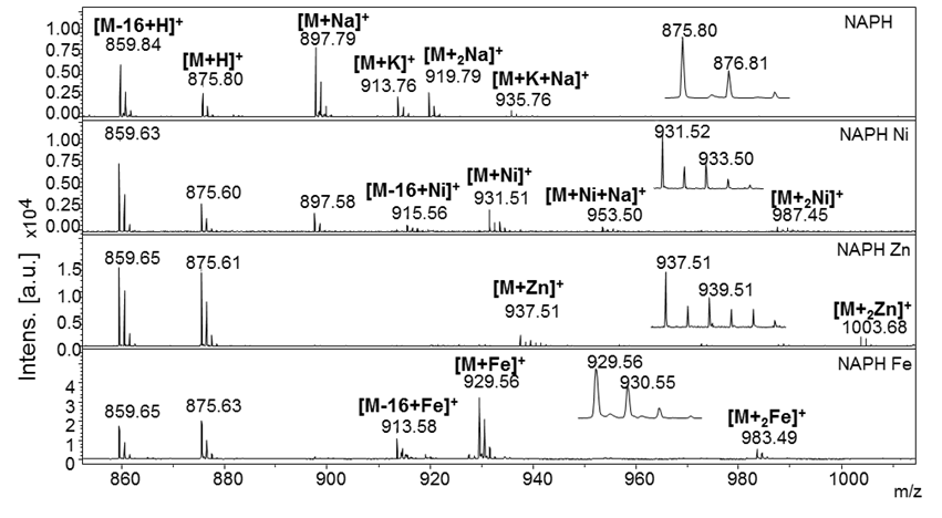
Figura 32. MS spectra of NAPH peptide (NH2-NAPVHIPQ-COOH) in the absence and presence of Ni2+, Zn2+ and Fe3+ ions at a molar ratio of 1:10 peptide: metal ion. The insertion in the spectrum corresponds to the isotopic distribution of the peak of interest.
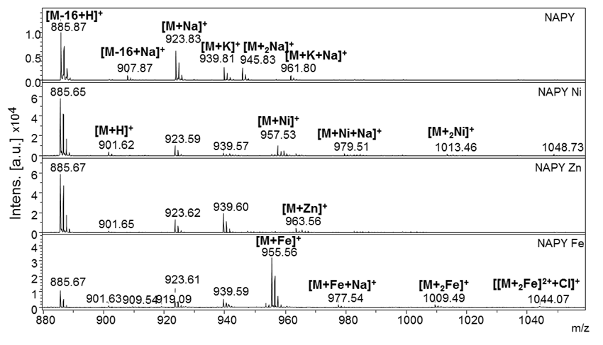
Fig. 33. MS spectra of the NAPY peptide (NH2-NAPVYIPQ-COOH) in the absence and presence of Ni2+, Zn2+ and Fe3+ ions at a molar ratio of 1:10 peptide: metal ion. The insertion in the spectrum corresponds to the isotopic distribution of the peak of interest.
II.2.12. Activity 3.1.
Characterization of complex systems involved in neurodegeneration by improved methods MS, FT-IR, NMR, AFM, DLS
The characterization of the amyloid-neuroprotective complex systems was achieved mainly by mass spectrometry, FT-IR and NMR, although some important data were obtained by DLS and AFM. In the works published with the participation of the doctoral students the MS and FT-IR data were presented, which can be easily accessed from the mentioned works. Therefore, we will present only general conclusions in this report. Thus, aluminum is an element that binds strongly to both amyloid and neuroprotective peptides.
II.2.13. Activity 3.2.
Study of the degradation of amyloid complexes
Systems consisting of amyloid peptides and neuroprotective NAP peptides have been developed that have been studied by mass spectrometry, FT-IR, NMR, etc. These studies have led to interesting results, namely that resveratrol may alter the degree of aggregation and conformation of amyloid peptides, which is the subject of a work being held at SGEM2019 in Albena, Bulgaria (Ref. 12: Ion, L., Drochioiu, G., Lupaescu, A., Jureschi, M., Petre, B. A. Amyloid-β and anti-amyloid peptides involved in Alzheimer`s disease: Interactions with metal ions. 19th International Multidisciplinary GeoConference SGEM2019, Albena, Bulgaria, 19(6.1) 515-522).
Other results are accepted at Revista de Chimee, September 2019. The results will be presented in detail in the final report.
II.2.14. Activity 3.3.
International and national conferences
In 2019 some of the project data was disseminated as well by PhD students and researchers who are members of the project at international conferences, such as SGEM2019, Albena, Bulgaria and others that will be widely reported in the final report
II.2.15. Activity 3.4.
Dissemination and patent application of the new investigation method by mass spectrometry
In the last year 2019 only 8 (eight) scientific papers in impact factor journals were published, other 2 (two) being accepted and will be published during this year or in the first months of 2020. Other 4 (four) scientific papers were presented at international conferences and published in ISI quoted volumes. All these works mentioned the financing from this project, Contract 56/2017. There have been researches regarding the patenting of some results, but many data have already been published, because the doctoral students wanted to complete the theses this year, according to the doctoral school program and they introduced the data in the theses and in the published works. That is why we are in the process of re-evaluating the remaining data that have not yet been published in order to patent them.
II.2.16. Activity 3.5.
Updating the web page
The project data and how to achieve its objectives have been constantly updated online.
III. Conclusions
The project plan was continuously followed by the project members and all its objectives and activities were fully realized. An HPLC instrument and the specific columns for peptides were purchased and the personnel involved in the project were trained. The materials and reagents needed to perform the proposed research in very good conditions were also purchased. Peptides such as A(4-10), A(1-10), A(1-28) and some cysteine and alanine analogs of the NAP peptide were synthesized and characterized. Peptides such as A(9-16) and its analogues with glycine and phenylalanine have been synthesized and characterized, by replacing amino acid residues of tyrosine and histidine, respectively. At the same time, the complexes of these peptides with metal ions such as zinc, copper or silver have been studied.
Five amyloid peptides were synthesized, purified and characterized: the native fragment Aβ (9-16), then two mutants, Aβ(9-16)Y10G, Aβ(9-16)Y10F in which tyrosine (Tyr, Y) from position 10 was modified with glycine (Gly, G) and phenylalanine (Phe, F). Two other mutant peptides, Aβ(9-16)H13GH14G, Aβ(9-16)Y10GH13GH14G, respectively, in which the two histidines (His, H) with glycine (Gly, G) and tyrosine (Tyr, Y) at position 10 with glycine (Gly, G) have been produced. At the same time, the complexes of these peptides with metal ions such as aluminum, iron, nickel or zinc have been studied. In the research, five new peptides derived from the NAPVSIPQ octapeptide were synthesized. Their synthesis was performed manually, using the solid phase technique according to the Fmoc/t-Bu strategy. The obtained compounds were then structurally characterized using MALDI-ToF mass spectrometry.
The synthetic peptides (NAPG, NAPA, NAPH and NAPY) can lead to understanding of the conformational changes of the native peptide, while the mutant with acetyl salicylic acid can be used as a multifunctional peptide, but also as a peptide-conjugated drug. Asp-NAP can be a peptide derivative with potential in reducing neuroinflammation and amyloid plaque formation. In this context, further research is required to determine its neuroprotective activity. The purity of the synthesized peptides was determined by high performance liquid chromatography (HPLC). As observed in their chromatograms, the peptides showed a single major signal corresponding to the pure peptide. The interaction studies between them with different metal ions were performed in the presence of nickel, zinc and iron metal ions at pH 7. Following the analysis of the obtained mass spectra, the formation of peptide-metal complexes was observed.
The interactions of amyloid peptides with neuroprotective ones, as well as with other anti-aggregating compounds, such as resveratrol, curcumin, etc. have been studied. And these results have already been presented and published like the other results in the project (at the international conference SGEM2019, Albena, Bulgaria).
Diseminarea rezultatelor s-a realizat şi prin prezentarea unui număr de 11 (unsprezece) lucrări ştiinţifice şi contribuţii, oral sau sub formă de postere la conferinţe internaţionale şi naţionale. Au fost redactate un număr de 15 (cinsprezece) lucrări ştiinţifice cu menţionarea finanţării din proiect. Astfel, au fost deja publicate 8 (opt) lucrări în jurnale cu factor de impact, alte două fiind acceptate la publicare, iar una a fost realizată şi trimisă la publicare în Journal of Peptide Science, o revistă cu factor de impact. Incă 4 (patru) lucrări au fost publicate în volumele conferinţelor internaţionale cotate ISI. Toate lucrările publicate, susținute sau trimise la publicare au conţinut un Acknowledgment cu nominalizarea finanţatorului şi a proiectului de idei, PN-III-P4-ID-PCE-2016-0376, Contract 56/2017.
The dissemination of the results was made by presenting a number of 11 (eleven) scientific papers and contributions, orally or as posters at international and national conferences. A number of 15 (fifteen) scientific papers were drafted mentioning the financing of the project. Thus, 8 (eight) papers in impact factor journals have already been published, two more being accepted for publication, and one has been published and published in the Journal of Peptide Science, an impact factor journal. Another 4 (four) papers were published in the volumes of the ISI indexed international conferences. All the works published, accepted or submitted to publication contained an Acknowledgment with the nomination of the financier and the ideas project, PN-III-P4-ID-PCE-2016-0376, Contract 56/2017.
IV. References
1. Cleary, J.P., Walsh, D.M., Hofmeister, J.J., Shankar, G.M., Kuskowski, M.A., Selkoe, D.J.
and Ashe, K.H., 2005. Natural oligomers of the amyloid-β protein specifically disrupt
cognitive function. Nature neuroscience, 8(1), 79-84.
2. Cummings, J. L. and Kaufer, D. Neuropsychiatric aspects of Alzheimer's diseas: The
cholinergic hypothesis revisited. Neurology, 47, 876-883, 1996.
3. Kang, J. E., Lim, M. M., Bateman, R. J., Lee, J. J., Smyth, L. P., Cirrito, J. R., Fujiki, N.,
Nishino, S., Holtzman, D. M. Amyloid-β dynamics are regulated by orexin and the sleep-wake cycle. Science, 326, 1005 – 1007, 2009.
4. Iwatsubo, T., Odaka, A., Suzuki, N., Mizusawa, H., Nukina, N. and Ihara, Y., 1994.
Visualization of Aβ42 (43) and Aβ40 in senile plaques with end-specific Aβ
monoclonals: evidence that an initially deposited species is Aβ42 (43). Neuron, 13(1),
45-53.
5. Haass, C. and Selkoe, D.J., 2007. Soluble protein oligomers in neurodegeneration: lessons
from the Alzheimer's amyloid β-peptide. Nature reviews Molecular cell biology, 8(2),
101-112.
6. Lesné, S., Koh, M.T., Kotilinek, L., Kayed, R., Glabe, C.G., Yang, A., Gallagher, M.
Ashe, K.H., 2006. A specific amyloid-β protein assembly in the brain impairs memory.
Nature, 440(7082), 352-357.
7. Kong, G.K.W., Adams, J.J., Harris, H.H., Boas, J.F., Curtain, C.C., Galatis, D., Masters, C.L.,
Barnham, K.J., McKinstry, W.J., Cappai, R. and Parker, M.W., 2007. Structural studies
of the Alzheimer’s amyloid precursor protein copper-binding domain reveal how it binds
copper ions. Journal of molecular biology, 367(1), pp.148-161.
8. Gozes I, Divinski I, Piltzer I, NAP and D-SAL: neuroprotection against the beta amyloid
peptide (1-42). BMC Neurosci. 9 Suppl 3, S3, 2008.
9. Gozes I, Morimoto BH, Tiong J, Fox A, Sutherland K, Dangoor D, Holser-Cochav M, Vered
K, Newton P, Aisen PS, Matsuoka Y, Dyck CH, Thal L, NAP: Research and
Development of a Peptide Derived from Activity-Dependent Neuroprotective Protein
(ADNP). CNS Drug Rev. 11, 353–368, 2006.
10. Magen I, Gozes I, Davunetide: Peptide therapeutic in neurological disorders. Curr. Med.
Chem. 21, 2591–8, 2014.
11. Kim, A., Lim, S., & Kim, Y. (2018). Metal Ion Effects on Aβ and Tau Aggregation.
International journal of molecular sciences, 19(1), 128.
12. Atrián-Blasco, E., Gonzalez, P., Santoro, A., Alies, B., Faller, P., & Hureau, C. (2018). Cu
and Zn coordination to amyloid peptides: From fascinating chemistry to debated
pathological relevance. Coordination chemistry reviews, 371, 38-55.
13. Liu, H., Qu, Y., & Wang, X. (2018). Amyloid β-targeted metal complexes for potential
applications in Alzheimer's disease. Future medicinal chemistry, 10(6), 679-701.
14. Gongora-Benitez, M., Mendive-Tapia, L., Ramos-Tomillero, I., Breman, A. C., Tulla-Puche,
J., & Albericio, F. (2012). Acid-Labile Cys-Protecting Groups for the Fmoc/t Bu
Strategy: Filling the Gap. Organic letters, 14(21), 5472-5475.
15. Kimmerlin, T., & Seebach, D. (2005). ‘100 years of peptide synthesis’: ligation methods for
peptide and protein synthesis with applications to β‐peptide assemblies. The Journal of
peptide research, 65(2), 229-260.
16. Oz, S. et al., 2014. The NAP motif of activity-dependent neuroprotective protein (ADNP)
regulates dendritic spines through microtubule end binding proteins. Molecular
Psychiatry, 19(10), 1115–1124.
17. Sarver, A. et al., 2001. Analysis of peptides and proteins containing nitrotyrosine by matrix-
assisted laser desorption/ionization mass spectrometry. Journal of the American Society
for Mass Spectrometry, 12(4), 439–448.
|